How to perform Otoscopy
What is otoscopy?
Before conducting audiometry, tympanometry or any other procedure on the ear, a clinician or healthcare professional will often conduct otoscopy. Otoscopy is a clinical procedure used to examine structures of the ear, particularly the External Auditory Canal (EAC), Tympanic Membrane ™, and to a lesser degree – provide an indication of the status of the Middle Ear (ME). Clinicians use an otoscope during routine wellness physical exams and the evaluation of specific ear complaints.
What is an otoscope?
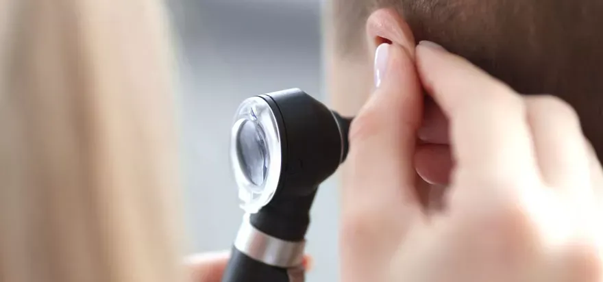
The otoscope consists of a light, a magnifying lens, and a funnel-shaped viewing piece with a narrow, pointed end called a speculum. The light beam is shined through the otoscope into the ear canal.
Otoscopy can be performed via manual or digital otoscopes. A manual otoscope can be used alone as the optic and the light source are within one handheld unit. A digital otoscope still provides the components in a handheld unit, but the image of the ear is displayed on a screen. This has additional benefits as it allows the user to photograph and document the otoscopy, or even show the image live to the patient.
Why is otoscopy important?
In an ideal test scenario, the ear canal is free and unblocked to allow acoustic energy (sound) and applied air pressure to transmit to the tympanic membrane and through the middle ear to the inner ear.
In the testing environments mentioned, ‘an ideal’ scenario is not always the case. Obstacles can be found in the ear canal, like ear wax build-ups, skin debris, fluids such as pus or foreign bodies.
Otoscopy not only helps to check that the path to the eardrum is clear, but also gives an excellent overview of the general condition of the ear canal and tympanic membrane to identify abnormalities such as a perforated eardrum, middle ear effusion, swelling or redness of the ear canal, infection or others.
How is otoscopy performed?
Otoscopy is performed by inserting the speculum into the patient’s ear and moving it in different directions to observe the condition of the EAC and TM. The clinician will gently pull the pinna upward and backward to straighten the ear canal.
This process will move the acoustic meatus in line with the canal to make it easier to observe landmarks within the ear and the overall health condition of the EAC and TM. This is a quick and painless process and an essential part of an auditory examination. Here are the steps to perform otoscopy:
- Prepare the equipment: Ensure that the otoscope is clean and has a working light source. If necessary, attach a disposable speculum to the otoscope
- Position the patient: Ask the patient to sit upright and tilt their head slightly towards the opposite shoulder to expose the ear canal
- Inspect the external ear: Use your fingers or the otoscope to inspect the external ear for any signs of inflammation, discharge, or foreign objects
- Insert the otoscope: Hold the otoscope with your dominant hand and gently insert the speculum into the ear canal while using your other hand to stabilise the patient’s head. Be careful not to insert the otoscope too deeply or forcefully
- Observe the ear canal and eardrum: Use the otoscope’s magnifying lens and light source to visualise the ear canal and eardrum. Look for any signs of redness, swelling, discharge, or abnormalities
- Remove the otoscope: When you are finished examining the ear, gently remove the otoscope from the ear canal
- Repeat the process: If you need to examine the other ear, repeat the same process on the other side.
It is important to be gentle and patient during the otoscopy procedure to avoid causing any discomfort or injury to the patient.